

Found a good "Pestilence & Plague" link? Let Us Know!
Some of the more prevalent varieties of P&P. Decontamination and treatment protocols, as well as methods of protecting against infection, continued on page 2.
| This Page: | All Topics/Misc. | Anthrax | Bird Flu | Botulinum |
| Cholera & Dysentery | Flesh Eating Bacteria | Hemorrhagic Fever | Other Bacterial | Pfiesteria |
| Plague | SARs | Smallpox | Tuberculosis | Tularemia |
| Next Page: | West Nile | (Other) Mosquito-Borne | Non-Plague Rodent | (Other) Tick Borne |
| Other Viruses | Antibiotics | Vaccines | Air Filters/Shelters | Protective Gear |
| Detection & Alarms | Clean-Up/DeCon | Threat Assessment | Treatment Protocols | Supplies/Suppliers |
All Topics/Misc.
The California Department of Health Services (CDHS) Pandemic Influenza
Preparedness and Response Plan: The plan outlines the roles and strategies of
CDHS in coordinating the public health response to a pandemic with local health
departments, the healthcare community, the federal government, and other key
partners. Scarey.
Infectious Diseases Emerging and Re-emerging Infectious Diseases. New
microorganisms capable of causing disease in humans continue to be detected
(see examples in Table 1). Whether an emerging microorganism develops into a
public health threat depends on factors related to the microorganism and its
environment, or the infected human and his/her environment. Such factors
include ease of transmission between animals and people and among people,
potential for spread beyond the immediate outbreak site, severity of
illness, availability of effective tools to prevent and control the
outbreak, and ability to treat the disease. Some of the new agents detected
in the past 25 years are now genuine public health problems on a local,
regional or global scale.
Epidemics
& Plagues: An epidemic is generally a widespread disease that affects many individuals in a population. An epidemic may be restricted to one locale or may even be global (pandemic). An outbreak of a disease is defined as being epidemic, however, not by how many members or what proportion of the population it infects but by how fast it is growing. When each infected individual is infecting more than one other individual, so that the number of infected individuals is growing exponentially, the disease is in an epidemic state. Thus even if the number of people affected is small, the phenomenon may still be called an epidemic, although for small epidemics the term "outbreak" is more often used.
Video:
"The History of Bioterrorism"
Describes the role of Category A agents—such as plague —as weapons
of bioterrorism
Words
of Wisdom About Gas, Germs, and Nukes By
SFC Red Thomas, Armor Master Gunner - U.S. Army (Ret) 10.19.01. Since the
media have decided to scare everyone with predictions of chemical, biological,
or nuclear warfare on our turf I decided to write a paper and keep things in
their proper perspective. I am a retired military weapons, munitions, and
training expert.
| Biological Agent Fact Sheet Info These fact sheets review the public health and medical consequences of selected biological agents deployed as weapons in a civilian community. They offer the consensus recommendations of The Johns Hopkins Working Group on Civilian Biodefense regarding appropriate medical and public health measures to be taken following such an attack. There are a number of candidate organisms terrorists could weaponize, but the Working Group identifies only a few that are widely known and feared and that would cause disease and deaths in sufficient numbers to cripple a city. | |||||
| Anthrax | Botulinum Toxin | Plague | Smallpox | Tularemia | VHF |
Bio-Terror
Survival Pretty good article
centering on anthrax and small pox. Cheesy site, too much advertising, but no
pop-ups.
United
States Army Medical Research Institute of Infectious Diseases (USAMRIID):
U.S.
Department of State Fact Sheets on
Chemical-Biological Warfare
Nunn-Lugar-Domenici
Domestic Preparedness & WMD Civil Support Teams
The Nunn-Lugar-Domenici Domestic Preparedness Program began in FY97 to train
first responders -- fire, police, and emergency medical technicians -- in 120 of
the largest cities in the country. Recently, the 120 cities that were designated
recipients of Nunn-Lugar-Domenici Domestic Preparedness Program have been
expanded and amended to a mixture of 157 cities and counties to replace the
original 120 cities.
USFA
- HAZMAT Guide for First Responders,
1999 The resources on this page will assist First Responders in safely
responding to a hazardous materials incident. Resources are also provided for
personnel who will assume the Incident Commander role in hazardous materials
emergencies above the initial response.
Protection
Against Chemical Weapons OPCW
Fact File based on "A FOA Briefing Book on Chemical Weapons"
Dr.
Kanatjan Alibekov (Ken Alibek) "Plague
War" on PBS's Frontline, aired October 13, 1998
Biotechnology
and Genetic Engineering: Implications for the Development of New Warfare Agents
1996 Office of Counterproliferation and
Chemical and Biological Defense, Office of the Secretary of Defense
The
Wonderful World of Diseases website
is about more than just diseases: it covers many other "human
conditions" that plague and intrigue us.
Identifying
Chemical and Biological Agents Learn the characteristics of these
substances and their effects. Chemical/biological agents are defined as
toxic substances, pathogens or toxins that are used to kill or incapacitate
people, animals or plants, or to damage equipment. The use of these agents dates
as far back as 429 B.C. when the Spartans ignited pitch and sulphur creating
toxic vapors in the Peloponnesian War. This article discusses the types of
chemical and biological agents and their effects. See also:
Chemical
Agents Chart (.pdf) requires Adobe Acrobat.
Tutorial: Chemical and Biological Agents
Chemical and biological (CB) agents pose
a threat to U.S. and allied military forces. Unfortunately, what strictly
used to be a military risk has now become a concern to the civilian
population—brought about by terrorist groups and other factions with
developed CB capabilities. As a result, national, state and local
authorities have initiated domestic preparedness against CB
terrorism. Although the probability of a terrorist CB attack is low, it’s
important to have an understanding of the potentially devastating and
far-reaching effects of these agents.
Meningococcal Epidemic Meningococcal Disease. Meningococcal meningitis
is characterized by sudden onset of intense headache, fever, nausea,
vomiting, photophobia, and stiff neck. Neurological signs include lethargy,
delirium, coma and/or convulsions. Infants may have illness without sudden
onset and stiff neck. Even when the disease is diagnosed early and adequate
therapy instituted, the case fatality rate is between 5% and 10% and may
exceed 50% in the absence of treatment. In addition to the mortality
associated with meningococcal meningitis 15% and 20% of those who survive
will suffer with neurological sequelae (e.g. deafness, mental retardation)
as a result of their illness. A less common but more severe (often fatal)
form of meningococcal disease is meningococcal septicemia which is
characterized by rapid circulatory collapse and a hemorrhagic rash.
Back to Top
Anthrax
Anthrax
& Anthrax
Vaccine CDC pages with tons of info...
HOW
TO HANDLE ANTHRAX AND OTHER BIOLOGICAL AGENT THREATS - CDC.gov Many facilities in communities
around the country have received anthrax threat letters.
Most were empty envelopes; some have contained powdery substances.
The purpose of these guidelines is to recommend procedures for handling
such incidents.
Clinical
and Epidemiologic Principles of Anthrax Theodore
J. Cieslak and Edward M. Eitzen, Jr., Journal of Emerging Infectious Diseases,
Vol. 5, No. 4 (July-Aug. 1999)
Anthrax:
A Possible Case History Thomas V. Inglesby, Journal of Emerging Infectious Diseases, Vol. 5, No. 4
(July-Aug. 1999)
Anthrax
as a Weapon of Terrorism and Difficulties Presented in Response to its Use
Defence Journal, December 1998
The
Official DOD Anthrax Information Website Department
of Defense, Information on troop vaccination, anthrax the disease, and anthrax
the threat.
Clinical
Anthrax: Primer for Physicians
Photo
of Cutaneous Anthrax on JAMA website
Anthrax
as a Biological Weapon, 2002: Updated Recommendations for Management
[JAMA 287(17),2002]
Anthrax
as a Biological Weapon: Medical & Public Health Management (1999)
[JAMA 281(18),1999]
Anthrax
as a Biological Weapon: Medical & Public Health Management by
Thomas V. Inglesby, Donald A. Henderson, John G. Bartlett, Michael S. Ascher,
Edward Eitzen, Arthur M. Friedlander, Jerome Hauer, Joseph McDade, Michael T.
Osterholm, Tara O'Toole, Gerald Parker, Trish M. Perl, Philip K. Russell, Kevin
Tonat for the Working Group on Civilian Biodefense JAMA. May 12, 1999;
281(18) The first in a series of six consensus statements published by the
Working Group on Civilian Biodefense outlining protocols for dealing with the
deliberate release of an infectious pathogen.
Anthrax
for the Pulmonary Physician by John Bartlett, Thomas V. Inglesby, Luciana Borio Pulmonary and
Critical Care Update-Online;16: Lesson 1. 2002 Online, didactic
article intended for pulmonary and critical care physicians. The article
addresses the epidemiology of anthrax, the diagnostic evaluation and management
of inhalational anthrax, and the rationale for antibiotic selection and
duration.
Anthrax
Awareness and Precautions Bacillus Anthracis are single-cell
organisms that cause anthrax. A bacterial disease, anthrax occurs in
domesticated and wild animals, including goats, sheep, cattle, horses and deer.
For anthrax to be effective as a covert agent, it must be aerosolized into very
small particles. This is difficult to do, and requires a great deal of technical
skill and special equipment. If these small particles are inhaled,
life-threatening lung infection can occur, but prompt recognition and treatment
are effective. See also:
Centers
for Disease Control and Prevention (CDC) FAQ's on Anthrax and
Anthrax
FAQ's
Back to Top
Bird Flu
Bird Flu & Avian influenza
Avian influenza (also known as bird flu) is a type of influenza virulent in birds. It was first identified in Italy in the early 1900s and is now known to exist worldwide.
Back to Top
Botulinum
The
Pathogenic Clostridia © 2002 Kenneth Todar
University of Wisconsin-Madison Department of Bacteriology: The clostridia are
ancient organisms that live in virtually all of the anaerobic habitats of nature
where organic compounds are present, including soils, aquatic sediments and the
intestinal tracts of animals. Most of the clostridia are saprophytes but a few
are pathogenic for humans. Those that are pathogens have primarily a saprophytic
existence in nature and, in a sense, are opportunistic pathogens. Clostridium
tetani and Clostridium botulinum produce the most potent biological
toxins known to affect humans. As pathogens of tetanus and food-borne botulism,
they owe their virulence almost entirely to their toxigenicity. Other
clostridia, however, are highly invasive under certain circumstances.
Positive
test for terror toxins in Iraq SARGAT, Iraq, April
4 — MSNBC.com tests reveal evidence of the deadly toxins ricin
and botulinum at a laboratory in a remote mountain region of northern Iraq
allegedly used as a terrorist training camp by Islamic militants with ties to
the al-Qaida terrorist network. The U.S. Central Intelligence Agency is
conducting its own tests at the same area, but has not yet released the results,
according to officials in northern Iraq.
Is
nation ready for botulinum attack? Experts fear trouble ahead
(CNN.com) WASHINGTON (AP) -- Easy to find
and easy to produce, botulinum toxin is the most poisonous natural substance on
Earth. In the hands of a bioterrorist, a single gram -- the weight of a paper
clip -- could kill more than 1 million people.
Botulinum
Toxin: From Poison to Medicinal Agent by
Jiri Patocka and Miroslav Splino. Botulinum toxin is very strong poison produced
by the microorganism Clostridium botulinum. C. botulinum is classified as a
single species but consists of at least three genetically distinguishable groups
of organisms. These are alike in their abilities to produce neurotoxins with
similar pharmacological activities [1] but diverse serologic properties (toxin
types A, B, C, D, E, F, and G). These types are defined by the International
Standards for Clostridium botulinum Antitoxin [2]. Botulinum toxins are the
causative agents of botulism, a potentially fatal condition of neuromuscular
paralysis. Botulism is characterized by symmetric, descending, flaccid paralysis
of motor and autonomic nerves, usually beginning with the cranial nerves.
Blurred vision, dysphagia, and dysarthria are common initial complaints. The
diagnosis of botulism is based on compatible clinical findings; history of
exposure to suspect foods; and supportive ancillary testing to rule out other
causes of neurological dysfunction that mimic botulism, such as stroke,
Guillain-Barré syndrome, and myasthenia gravis. Treatment includes supportive
care and trivalent equine antitoxin, which reduces mortality if administered
early.
Botulinum
Toxin Fact Sheet Botulinum toxin poses a major bioweapons threat because of
its extreme potency and lethality; its ease of production, transport and
misuse; and the potential need for prolonged intensive care in affected
persons. Botulinum toxin is the single most poisonous substance known.
Botulinum
Toxin as a Biological Weapon: Medical & Public Health Management
An aerosolized or foodborne botulinum toxinweapon would cause acute
symmetric, descending flaccid paralysiswith prominent bulbar palsies
such as diplopia, dysarthria,dysphonia, and dysphagia that would
typically present 12 to72 hours after exposure. Effective response
to a deliberaterelease of botulinum toxin will depend on timely
clinical diagnosis,case reporting, and epidemiological
investigation. Persons potentiallyexposed to botulinum toxin should
be closely observed, and thosewith signs of botulism
require prompt treatment with antitoxinand supportive care that may
include assisted ventilation forweeks or months. Treatment with
antitoxin should not be delayedfor microbiological testing.
Clostridium
botulinum is an anaerobic,
Gram-positive,
spore-forming rod that produces a potent
neurotoxin.
The
spores
are heat-resistant and can survive in foods that are incorrectly or minimally
processed. Seven types (A, B, C, D, E, F and G) of
botulism
are recognized, based on the antigenic specificity of the toxin produced by each
strain. Types A, B, E and F cause human botulism. Types C and D cause most cases
of botulism in animals. Animals most commonly affected are wild fowl and
poultry, cattle, horses and some species of fish. Although type G has been
isolated from soil in Argentina, no outbreaks involving it have been recognized.
Microbiology
Laboratory Guidebook 3rd
Edition/1998. (PDF) CHAPTER 14. METHODS
FOR THE DETECTION OF CLOSTRIDIUM BOTULINUM. TOXINS IN MEAT AND POULTRY
PRODUCTS. L. Victor Cook.
Back to Top
Cholera & Dysentery
Cholera Cholera is an acute intestinal infection caused by the bacterium
Vibrio cholerae. It has a short incubation period, from less than one day to
five days, and produces an enterotoxin that causes a copious, painless,
watery diarrhea that can quickly lead to severe dehydration and death if
treatment is not promptly given. Vomiting also occurs in most patients. Most
persons infected with V. cholerae do not become ill, although the bacterium
is present in their feces for 7-14 days. When illness does occur, more than
90% of episodes are of mild or moderate severity and are difficult to
distinguish clinically from other types of acute diarrhea. Less than 10% of
ill persons develop typical cholera with signs of moderate or severe
dehydration.
Dysentery Dysentery may be simply defined as diarrhea containing blood.
Although several organisms can cause dysentery, Shigella are the most
important. Shigella dysenteriae type 1 (Sd1), also known as the Shiga
bacillus, is the most virulent of the four serogroups of Shigella. Sd1 is
the only cause of epidemic dysentery. In addition to bloody diarrhea, the
illness caused by Sd1 often includes abdominal cramps, fever and rectal
pain. Less frequent complications of infection with Sd1 include sepsis,
seizures, renal failure and the haemolytic uraemic syndrome. Approximately
5-15% of Sd1 cases are fatal.
Flesh Eating Bacteria
Flesh-Eating
Bacteria: Facts Behind the Bug Necrotizing
fasciitis is a bacterial infection. These bacteria attack the soft tissue,
usually in an extremity following minor trauma. There are also many cases of
this occurring after surgery, and most often abdominal surgery. The name
"flesh-eating-bacteria" is a little sensational, but essentially, the
bacteria do "eat flesh." They attack the subcutaneous (soft) tissue,
which then becomes gangrenous. Infection moves swiftly, usually under the skin,
where it is unobservable. Once tissue becomes necrotic (dead), it has to be
removed.
Necrotizing Fasciitis: Pathophysiology and Treatment by Anne Fink BSN, RN and Gail Deluca MS, RN-c, CNP Medical-Surgical Nursing Journal
- Online CE
Necrotizing Fasciitis
This is a brief, but very informative fact sheet.
Necrotizing Fasciitis Clinical Resources A very thorough index of articles from the
University of Alabama, which requires a registration with a login and password.
Severe Invasive Group A Streptococcal Infections: A Subject Review (RE9804) American Academy of Pediatrics
Outbreak of Invasive Group A Streptococcus Associated with Varicella in a Childcare Center -- Boston, Massachusetts, 1997
Avoiding
Contagion with Necrotizing Fasciitis Dr. Harold Oster, ivillagehealth.com
Group A Streptococcal (GAS) Disease
Invasive
Group A Strep New York State Department of Health Communicable Disease Fact
Sheet
Group
A Streptococcal Infections National Institute of Allergy and Infectious
Diseases
Epidemiologic
Notes and Reports Nursing Home Outbreaks of Invasive Group A Streptococcal
Infections -- Illinois, Kansas, North Carolina, and Texas
Streptococcal
Toxic-Shock Syndrome: Spectrum of Disease, Pathogenesis, and New Concepts in
Treatment
Nosocomial
Group A Streptococcal Infections Associated with Asymptomatic Health-Care
Workers -- Maryland and California, 1997
INVASIVE
GROUP A STREPTOCOCCUS IN TWO SIBLINGS: A CASE FOR ANTIBIOTIC PROPHYLAXIS OF
CLOSE CONTACTS Canadian Population and Public Health Branch
Role
of CT in Necrotizing Fasciitis of the Head and Neck
NECROTIZING
FASCIITIS OF THE HEAD AND NECK Troy A. Callender, MD December 31, 1992
Necrotizing
fasciitis after Plastibell circumcision
Hyperbaric
Oxygen Therapy In Necrotizing Fasciitis: Panacea, Useful Adjunct, or Nostrum?
Back to Top
Hemorrhagic Fever
Viral
Hemorrhagic Fevers Fact Sheet
(What are viral hemorrhagic fevers?)
Viral hemorrhagic fevers (VHFs) refer to a group of illnesses that are caused by
several distinct families of viruses. In general, the term "viral
hemorrhagic fever" is used to describe a severe multisystem syndrome (multisystem
in that multiple organ systems in the body are affected).
Characteristically, the overall vascular system is damaged, and the body's
ability to regulate itself is impaired. These symptoms are often
accompanied by hemorrhage (bleeding); however, the bleeding is itself rarely
life-threatening. While some types of hemorrhagic fever viruses can cause
relatively mild illnesses, many of these viruses cause severe, life-threatening
disease.
What
is Ebola hemorrhagic fever? Ebola
hemorrhagic fever (Ebola HF) is a severe, often-fatal disease in humans and
nonhuman primates (monkeys, gorillas, and chimpanzees) that has appeared
sporadically since its initial recognition in 1976.
List
of Known Cases and Outbreaks of Ebola Hemorrhagic Fever
Table Showing Known Cases and Outbreaks, in Chronological Order
Filoviruses
Fact Sheet (What are filoviruses?)
Filoviruses belong to a virus family called Filoviridae and can cause severe
hemorrhagic fever in humans and nonhuman primates. So far, only two members
of this virus family have been identified: Marburg virus and Ebola virus. Four
species of Ebola virus have been identified: Ivory Coast, Sudan, Zaire, and
Reston. Ebola-Reston is the only known filovirus that does not cause severe
disease in humans; however, it can be fatal in monkeys.
Infection
Control Manual In 1995, an outbreak of Ebola
hemorrhagic fever (Ebola HF) affected more than 300 people in and around the
city of Kikwit, Democratic Republic of the Congo (formerly, Zaire);
approximately 80% of the patients died. More than one-fourth of all the patients
were health care workers. After the outbreak, the DRC Ministry of Health, the
Centers for Disease Control and Prevention (CDC) and the World Health
Organization (WHO) developed practical recommendations for carrying out viral
hemorrhagic fever (VHF) isolation precautions in rural health facilities in
Africa. These recommendations have been consolidated in a manual for the local
health community.
Past
Outbreak Postings (since 2000) This
section has archived postings of outbreaks that have occured since the year
2000. For information on current outbreaks, please refer to the
Special
Pathogens home page. To find information on outbreaks that have occurred
prior to the year 2000, please visit the
Other
Resources page.
Marburg
Hemorrhagic Fever Fact Sheet Marburg
hemorrhagic fever is a rare, severe type of hemorrhagic fever which affects both
humans and non-human primates. Caused by a genetically unique zoonotic (that is,
animal-borne) RNA virus of the filovirus family, its recognition led to the
creation of this virus family. The four species of Ebola virus are the only
other known members of the filovirus family.
The
Hemorrhagic Fever Viruses as Biological Weapons: Medical & Public Health
Management [JAMA 287(18),2002]
The
Hemorrhagic Fever Viruses as Biological Weapons:Medical and Public Health
Management by Luciana Borio, Thomas
V. Inglesby, C.J. Peters, Alan L. Schmaljohn, James M. Hughes, Peter B. Jahrling,
Thomas Ksiazek, Karl Johnson, Andrea Meyerhoff, Tara O'Toole, Michael S. Ascher,
John Bartlett, Joel G. Breman, Edward Eitzen, Margaret Hamburg, Jerry Hauer,
Donald A. Henderson, Richard T. Johnson, Gigi Kwik, Marci Layton, Scott
Lillibridge, Gary J. Nabel, Michael T. Osterholm, Trish M. Perl, Philip K.
Russell, Kevin Tonat for the Working Group on Civilian Biodefense JAMA.
May 8, 2002; 287(18) Consensus recommendations from the Working Group on
Civilian Biodefense outlining protocols for dealing with a deliberate release of
viral hemorrhagic fever pathogens.
List
of VHFs that SPB works with
The Special Pathogens Branch works with Biosafety Level 4 (BSL-4)
viruses. These viruses are highly pathogenic and require handling in special
laboratory facilities designed to contain them. Our
mission
focuses mainly on viral hemorrhagic fevers (VHFs), such as Ebola hemorrhagic
fever, Lassa fever, hantavirus pulmonary syndrome (HPS), and hemorrhagic fever
with renal syndrome (HFRS). Our work also includes
the study of other emerging viral disease agents that do not cause hemorrhagic
fever, but require BSL-4 handling, such as Nipah virus and tick-borne
encephalitis virus.
Lassa
Fever Fact Sheet Lassa fever is an acute viral illness that occurs in
West Africa. The illness was discovered in 1969 when two missionary nurses died
in Nigeria, West Africa. The cause of the illness was found to be Lassa virus,
named after the town in Nigeria where the first cases originated. The virus, a
member of the virus family Arenaviridae, is a single-stranded RNA virus and is
zoonotic, or animal-borne. See also:
"Lassa"
Online Video &
Lassa
Fever Slideset
Back to Top
Other Bacterial
Escherichia coli is a bacterium that is a common inhabitant of the
gut of warm blooded animals, including man. Most strains of E. coli are
harmless, however, some strains, such as E. coli O157:H7, can cause severe
foodborne disease and are referred to as enterohaemorrhagic E .coli (EHEC).
This pathogen produces toxins, known as verotoxins or Shiga-like toxins
because of their similarity to the toxins produced by Shigella dysenteriae.
The organism can grow from around 7-10C to 50C, with an optimum temperature
of 37C. Some EHEC can grow in acidic foods, down to a pH of 4.4, and in
foods with a minimum water activity (Aw) of 0.95. It is destroyed by
thorough cooking of foods until all parts reach a temperature of 70C or
higher. The designation “O157:H7” in the name of this bacterium refers to
specific chemical compounds that are found on its surface that distinguishes
it from other strains of E. coli.
Salmonella MULTI-DRUG RESISTANT SALMONELLA TYPHIMURIUM: It was in 1885
that Daniel E. Salmon, U.S. veterinary surgeon, discovered the first strain
of Salmonella. Today, there are 2213 known strains and the book is not
closed. Over the years, antibiotic resistant strains have developed that are
difficult to control and there is a body of evidence in the scientific
literature suggesting the possibility that some of these strains may have
emerged due to use of antibiotics in intensive animal husbandry. Recent
years saw a dramatic rise both in terms of incidence and severity of cases
of human salmonellosis; compared to 1980 some countries in Europe witnessed
a staggering 20-fold increase in incidence in the last 10 - 15 years.
Typhoid Typhoid fever is contracted when people eat food or drink water
that has been infected with Salmonella typhi. It is recognized by the sudden
onset of sustained fever, severe headache, nausea and severe loss of
appetite. It is sometimes accompanied by hoarse cough and constipation or
diarrhea. Case-fatality rates of 10% can be reduced to less than 1% with
appropriate antibiotic therapy. Paratyphoid fever shows similar symptoms,
but tends to be milder and the case-fatality rate is much lower. The annual
occurrence of typhoid fever is estimated at 17 million cases, with
approximately 600,000 deaths. Some strains of Salmonella typhi are resistant
to antibiotics.
Pfiesteria
PFIESTERIA
HYSTERIA? - OR A REAL THREAT TO
MARINE LIFE? Not too long but very interesting article.
Pfiesteria
& Harmful Algal Blooms in the Mid-Atlantic.
Pfiesteria piscicida, a tiny marine organism identified in the last
decade in estuaries in North Carolina and Delaware and in the Chesapeake Bay and
its tributaries, has been blamed for killing fish and causing health problems in
humans. It has been the subject of national press coverage and the cause of
considerable confusion and speculation. While questions about its effects and
impacts remain, thanks to the diligent work of a number of marine scientists we
now know much more about this complicated organism.
Background
Information on Pfiesteria The
toxic dinoflagellate, Pfiesteria piscicida, has been implicated as the
primary causative agent of major fish kills and fish disease events (finfish and
shellfish; 10 to 10 finfish) in North Carolina estuaries, coastal areas, and aquaculture operations.
Pfiesteria and closely related toxic species ("Pfiesteria-like
complex") have also been confirmed in fish kill/disease areas and
aquaculture facilities outside North Carolina from the mid-Atlantic to the Gulf
Coast. Fish kills caused by P. piscicida usually occur in the warmest
part of the year, and often precede low dissolved oxygen levels in the
estuaries.
Pfiesteria
facts. Pfiesteria
piscicida (fee-STEER-ee-uh pis-kuh-SEED-uh), first discovered in 1988, occurs
naturally in the environment.
The
Cell from Hell, from Micscape
Magazine (in the UK), provides some thoughts on Pfiesteria.
The Chesapeake Bay Program has information about
Pfiesteria.
A Congressional Research Service report,
Pfiesteria
and Related Harmful Blooms: Natural Resource and Human Health Concerns
is available. The report examines the science of Pfiesteria and the
policy implications of Federal, state, and local responses.
Pfiesteria
piscicida / Pfiesteria shumwayae, from the Maryland Department of
Natural Resources covers these Pfiesteria-related topics: who's involved,
areas affected, chronology, who to contact; provides press releases, technical
information and fact sheets. Also provides access to various reports, including the
Report
of the Governor's Blue Ribbon Citizens Pfiesteria Action Commmission
(the Hughes Report).
Fish
Health, Fungal Infections, and Pfiesteria: The Role of the U.S. Geological
Survey (USGS) provides information on USGS investigations of Pfiesteria
in the Chesapeake Bay region. Also see a related
press
release (9/26/97).
Microscopic
Murderer: Pollution May be Motivating Pfiesteria to Kill Fish by the
Thousands is a new environmental fact sheet produced by the Academy of
Natural Sciences (August 1998).
No
Hysteria Over Pfiesteria is information from Pamlico County,
North Carolina.
The North Carolina Department of Environment and Natural Resources offers a
question
and answer fact sheet on Pfiesteria. There's also a
glossary
of terms related to the topic.
Pfiesteria
and Agriculture, from the University of Maryland College of Agriculture
and Natural Resources, provides information on the topic, including a white
paper (revised November 1997)
(HTML version)
(PDF
version) developed by the college.
Pfiesteria
piscicida Bibliography of the scientific literature was produced by
the Water Quality Information Center at the National Agricultural Library. Also,
many Pfiesteria piscicida articles are included in a
list
of publications provided by the North Carolina State University Center for
Applied Aquatic Ecology.
Surveillance
for Possible Estuary-Associated Syndrome---Six States, 1998--1999 is an
article in Morbidity and Mortality Weekly (May 05, 2000 / 49(17);372-3)
from the Centers for Disease Control and Prevention.
Pfiesteria
research information is provided by the Virginia Institute of Marine
Science. The Virginia Marine Resources Commission also offers links to
Pfiesteria
Information.
Back to Top
Plague
CDC
Plague Home Page Plague is an infectious disease of animals and humans
caused by a bacterium named Yersinia pestis. People
usually get plague from being bitten by a rodent flea that is carrying the
plague bacterium or by handling an infected animal. Millions of people in Europe
died from plague in the Middle Ages, when human homes and places of work were
inhabited by flea-infested rats. Today, modern antibiotics are effective against
plague, but if an infected person is not treated promptly, the disease is likely
to cause illness or death.
Plague
Manual: Epidemiology, Distribution, Surveillance & Control
Communicable Surveillance &
Response (CSR), World Health Organization. On their site
The
Black Plague (1348-1350) General
Information Primary Sources General Information: HWC, The Black Death : The BEST
source on the Plague (23 pages)
Q
& A About Plague Provided
by the Division of Vector-Borne Infectious Diseases, NCID, CDC
Consensus Statement: Plague as a Biological Weapon: Medical & Public Health Management
JAMA 2000;283(17):2281-2290.
Plague:
Diagnosis From the
CDC
Plague Home Page, provided by the Division of Vector-Borne Infectious
Diseases, NCID, CDC
Recognition
of Illness Associated with the Intentional Release of a Biologic Agent
MMWR 2001 Oct 19;50(41):893-897.
Facts
About Pneumonic Plague
FAQ
About Plague
Back to Top
SARs
WHO:
Severe Acute Respiratory Syndrome (SARS)
WHO:
SARS FAQ
CDC:
Severe Acute Respiratory Syndrome (SARS) Documents
Health Canada
Sars Site
HealthCanada:
Severe Acute Respiratory Syndrome
Health
Canada: Learning from SARS - Renewal of Public Health in Canada
BCGSC:
British Columbia Genome Sciences Centre - SARS Page [sequence data]
CMAJ:
Canadian Medical Association Journal on SARS
New
England Journal of Medicine: SARS articles
2003/03/12:
WHO: WHO issues a global alert about cases of atypical pneumonia
SLU:CSEI:
Centre for the Study of Emerging Infections on SARS
2003/03/15:
CDC: Severe Acute Respiratory Syndrome - CDC Issues Health Alert About Atypical
Pneumonia
MedLinePlus:
Atypical pneumonia
(website)
CNA: SARS Special
NewScientist:
SARS
Guardian(UK)
on SARS
CUNY:
Government Views of SARS
UVic:
SARS Bioinformatics Suite
SARS
Bioinformatics Suite
Province
of Manitoba Health Department on SARS
Toronto
Health Department on SARS
University
of Leicester: Coronaviruses & SARS
Softberry:
Annotated SARS genome sequence
The
Big Picture Book of Viruses: Coronaviridae
Algonomics:
List of SARS epitopes
Nature
web focus: SARS
SARS
Reference
Back to Top
Smallpox
Smallpox:
Clinical and Epidemiologic Features D.
A. Henderson, Journal of Emerging Infectious Diseases, Vol. 5, No. 4
(July-Aug. 1999)
Smallpox:
An Attack Scenario Tara O'Toole, Journal
of Emerging Infectious Diseases, Vol. 5, No. 4 (July-Aug. 1999)
Smallpox
as a Biological Weapon: Medical & Public Health Management
[JAMA 281(22),1999]
Smallpox:
An Attack Scenario by Tara O'Toole Emerging Infectious Diseases. Jul-Aug 1999; (5)4
Scenario conducted at the First National Symposium on the Medical and Public
Health Response to Bioterrorism, convened by the Center for Civilian Biodefense
Studies in Crystal City, Virginia on February 16-17, 1999.
Smallpox:
Clinical and Epidemiological Features by Donald A.
Henderson Emerging Infectious Diseases. Jul-Aug,1999; 5(4) Outlines,
for clinicians who have never come in contact with a case of smallpox, the
characteristics of the disease.
Smallpox
as a Biological Weapon: Medical & Public Health Management
by Donald A. Henderson, Thomas V. Inglesby, John G. Bartlett, Michael S. Ascher,
Edward Eitzen, Peter B. Jahrling, Jerome Hauer, Marcelle Layton, Joseph McDade,
Michael T. Osterholm, Tara O'Toole, Gerald Parker, Trish M. Perl, Philip K.
Russell, Kevin Tonat for the Working Group on Civilian Biodefense JAMA.
June 9, 1999; 281(22) Consensus recommendations from the Working Group on
Civilian Biodefense outline protocols for dealing with a deliberate release of
smallpox.
Smallpox
FAQ's 3 page .pdf file requires Adobe Acrobat.
Back to Top
Tuberculosis
Tuberculosis (TB) TB is a contagious disease. Like the common cold, it
spreads through the air. Only people who are sick with pulmonary TB are
infectious. When infectious people cough, sneeze, talk or spit, they propel TB
germs, known as bacilli, into the air. A person needs only to inhale a small
number of these to be infected. Left untreated, each person with active TB will
infect on average between 10 and 15 people every year. But people infected with
TB will not necessarily get sick with the disease. The immune system 'walls off'
the TB bacilli which, protected by a thick waxy coat, can lie dormant for years.
When someone's immune system is weakened, the chances of getting sick are
greater.
Tularemia
Tularemia
Fact Sheet Francisella tularensis, the organism that causes tularemia, is
one of the most infectious pathogenic bacteria known, requiring inoculation
or inhalation of as few as 10 organisms to cause disease. It is considered
to be a dangerous potential biological weapon because of its extreme
infectivity, ease of dissemination, and substantial capacity to cause
illness and death.
Tularemia
as a Biological Weapon: Medical & Public Health Management
[JAMA 285(21),2001]
Francisella
tularensis is considered a potential bioterrorism agent. An aerosol release
would be the most likely method and would result in a large number of
pneumonia cases several weeks after exposure.
Why
is there concern about tularemia as a bioweapon? Tularemia,
in aerosol form, is considered a possible bioterrorist agent. Persons who inhale
an infectious aerosol would likely experience severe respiratory illness. Any
suspected cases of tularemia inhalation should be immediately reported to local
and state health departments.
Tick-Borne
Diseases, Tularemia Although
numerous animals and insects can carry F tularensis, rabbits and ticks
(especially Dermacentor and Amblyomma species) most commonly
are implicated in human cases. The deer fly is another classic, although less
common, vector.
Back to Top

Hibiclens Antiseptic, Antimicrobial Skin Cleanser, Liquid
- 16 fl oz

N95 Respirators - (2 Masks) Approved - Pandemic Influenza Flu Protection
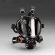
7000 Series Full Facepiece Respirators Full Face Respirator Medium

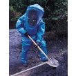
Tychem BR Hazmat Suit Chemical Protective Clothing W/ Attached Gloves &
Booties, Zipper Front Closure & Removable Hood. Includes Kevlar Glove
Liners.
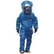
DuPont Tychem Responder Protective Apparel, Level B Suit
DuPont Tychem Responder Protective Apparel, Level A (Gas-Tight) Suit